All published articles of this journal are available on ScienceDirect.

The Skin–brain Axis in Cutaneous Aging: Neurobiological Foundations and Clinical Implications
 iD
Haitham Bader1
iD
Waseem H. Q. Alkhaffaf1
iD
Yahya D. M. Zakaria1
iD
Omar Y. Abdullah1
iD
Mohammed N. S. Al-Mallah1
iD
Authors Info & Affiliations
Abstract
The skin– brain axis is a bidirectional neuroimmunoendocrine axis connecting psychological stress, brain signaling, immune responses, and skin homeostasis. A hypothetical link between dysregulation of this network and skin ageing is neurogenic inflammation, oxidative stress, cellular senescence, inflammaging, and activation of the hypothalamic–pituitary–adrenal axis. This dysregulation is increasingly recognized as associated with mechanisms of skin ageing, including neurogenic inflammation, oxidative stress, cellular ageing, inflammaging, and activation of the hypothalamic–pituitary–adrenal axis. This review focuses on the neurobiological mechanisms underlying cutaneous ageing and emphasizes the role of neurocutaneous receptors, neurotransmitters, neuropeptides, and neuro-signaling pathways in the stress response. It also examines emerging trends, such as 'neurocosmetics' including peptides that act on neurocutaneous pathways to stimulate regeneration, stress-modulating peptides, and myorelaxant peptides. This review provides new insight into the skin–brain axis as a potential model for better understanding stress-related skin ageing by combining intrinsic and extrinsic skin ageing with neurobiology. There are a variety of neuroactive cosmetic strategies, with some having mechanistic and preliminary clinical evidence, while others do not and require controlled, randomized studies with objective markers. Future preventive and therapeutic strategies to regulate neuroimmune and neuroendocrine signaling to preserve skin homeostasis and reduce stress-induced skin ageing may be available.
1. INTRODUCTION
In medical practice, the skin and the nervous system are well known to be connected. However, this connection has only recently begun to shape new concepts in skin physiology. Today, discussion of the “cutaneous nervous system” centers on the skin as an organ of sensory and neuroimmune function that decodes emotional states and environmental signals [1]. This change has laid the foundation for therapeutic and cosmetic strategies that impact the skin's function and psychological state. The key aspect of this emerging field is that keratinocytes, melanocytes, and resident immune cells can both produce and respond to neurotransmitters, including endorphins, serotonin, dopamine, and substance P. These mediators help regulate barriers, modulate inflammation, and shape sensory perception. This creates a peripheral neuroendocrine network linked to emotional states and stress responses. This role may also help explain why emotional stress can worsen atopic dermatitis, psoriasis, and rosacea, all of which have been associated with disruption of neuroimmune signaling along the skin-brain axis [1, 2]. This relationship is increasingly understood as knowledge of skin microbiomes grows. A disturbance in microbial balance can activate systemic inflammatory pathways, which can influence Hypothalamic– Pituitary–Adrenal (HPA) activity and consequently affect affective regulation and skin inflammation [1, 3]. Recent studies also suggest that microbial metabolites and antibiotic exposure influence neuro-inflammatory responses that could underlie stress-related skin reactivity [4]. While researchers are still exploring the mechanisms behind these effects, microbiome-targeted topical approaches are gaining traction, with preliminary links established between skin composition and psychological and skin effects [4]. Meanwhile, the notion that topical products could affect emotional health adds complexity. Various agents have been suggested to modulate skin reactivity, increase comfort, and influence subjective sensations about skin condition, such as acetylhexapeptide-8, cannabinoid receptor agonists, and agents related to endorphin signaling [5]. Such statements are at least open to questioning because therapies affecting the neurosensory pathways could also influence emotional reactions. This matters because it is a biochemical effect that is not necessarily what one expects to see physiologically, and a physiologically measurable effect that may reflect a well-documented placebo effect [3-5].
Meanwhile, digital technologies are changing how we define this field. Through innovative artificial intelligence technologies, indicators of emotion can be captured from facial microexpressions, thermopatterns, and circadian or autonomic signals, enabling more personalized and responsive skin care [6]. These technologies can provide benefits but also raise data privacy and governance concerns, as well as other challenges in real-world care delivery. As these are developed, ethical issues need to be considered to ensure that they improve patients' health and do not create new dangers. Taken together, these trends suggest we may be seeing the beginning of a multidisciplinary field spanning dermatology, neuroscience, psychology, sensory biology, and data science. Much of the advancement is theoretical, and far more work is required for these ideas to have clinical impact. Standardized methods, objective parameters, and studies using longer periods are needed in the future, with integration of dermatological and psychophysiological parameters [1, 2]. If this groundwork is laid, emotionally responsive skin care may become a vital aspect of integrative skin care. This field focuses on the dynamic relationship between emotions, the brain, and skin [7]. This convergence is important for the discipline. It broadens the concept of skin as a barrier to an interface inhabited by neurological, immunological, microbial, and psychological processes. Future research on these cutaneous neural pathways, microbially influenced processes, and sensory function could lead to novel approaches to optimizing patient treatments, especially for patients whose skin conditions are closely linked to emotional fluctuations and neuroimmunological sensitivities [8].
Skin ageing is a complex process that involves both genetic ageing mechanisms (intrinsics) and environmental ageing mechanisms (extrinsics) [9]. Although UV exposure and oxidative stress are important, cumulative stress is also important. In recent years, it has been clearly demonstrated that the measurable contribution of psychological stress and interactions between the nervous system and immune system in skin homeostasis are of great importance [9]. This interaction has a developmental basis, and the shared embryologic origin of the epidermis and the nervous system underlies it [10]. Neuroimmunocutaneous models offer mechanistic insight into how emotional and physiological stress affects the aging phenotype [3-5]. Thus, the skin–brain axis has become an exciting field of study again, given its role in modulating skin resilience, inflammatory tone, and regeneration, following the integration of dermatology with neuroscience and immunology.
1.1. Study Design and Literature Search Strategy
This study aimed to conduct a narrative literature review to summarize existing knowledge on the mechanisms in the skin–brain axis involved in cutaneous aging and their possible clinical implications. We searched PubMed/MEDLINE, Scopus, Web of Science, and Google Scholar, focusing on peer-reviewed English-language literature. The following keywords were used in the search: “skin–brain axis,” “cutaneous aging,” “skin aging,” “neurogenic inflammation,” “neuroendocrine signaling,” “oxidative stress,” “inflammaging,” “cellular senescence,” “neuropeptides,” “neurotransmitters,” and “neurocosmetics.” We included original studies, reviews, and clinical and experimental reports relevant to the mechanisms, biomarkers, and potential therapeutic approaches of the skin–brain axis and aging. We also scanned relevant journals and newspapers to identify additional studies. We synthesized the literature narratively based on the key mechanistic and clinical themes discussed in this review.
2. HISTORY OF THE BRAIN–SKIN LINK
One of the oldest written texts that documents the skin- brain bond is found in the Charaka Samhita of ancient Indian medicine, “Ayurveda,” written in the 2nd century CE: the skin (Twacha) is closely linked to the mind (Manas), as described in the quoted Chetasamvayi (inseparable relation) [11]. A similar observation appears in ancient Greek medicine and Avicennian traditions, which recognize a deep, innate bond between the brain and skin, linking them through their shared embryological origin [12]. Hippocrates, in Epidemics III (410 BC), reported on the case of Thasos, the wife of Delearces [3]. Following a bout of grief, she had periodic fevers, wrapped herself up, and plucked, scratched, and picked at her hair [13]. Traditional Chinese Medicine views skin disorders as manifestations of internal imbalance. Practitioners use acupuncture and herbal remedies that target the nervous system to restore harmony [14]. In modern medicine, Psychodermatology has emerged as a discipline dedicated to exploring this complex relationship. It aims to comprehend the bilateral dialogue between the skin and the brain and how treating either alone can lead to incomplete management and early relapses [15].
3. CHARACTERISTICS OF THE SKIN–BRAIN AXIS
3.1. Common Embryonic Origin
The ectoderm contributes to the development of both the skin's epithelial layer and the nervous system [16]. During gastrulation, ectodermal cells, influenced by Wnt, FGF, and BMP signaling pathways, differentiate into either neural or epidermal cells [17]. FGF promotes neural cell formation, while blocking it induces BMP expression and commits ectodermal cells to become epidermal cells [18]. Later, the central neural plate in the ectoderm folds inward to form the neural tube, which develops into the central nervous system. In contrast, the peripheral ectoderm differentiates to form the skin and its appendages. Many molecular cues orchestrate epidermal specification, commitment, stratification, and terminal differentiation.
3.2. Heavy Neural Connection
Because of their shared embryonic development, the brain maintains a strong neural connection with the skin throughout life [19]. The skin is therefore not a simple passive barrier; rather, it is a smart dress for the body and represents its external boundary between the inside and the outside. Therefore, it is regarded as an “external” component of the nervous system [20]. It is estimated that the skin across the whole body is innervated by approximately 230,000 tactile afferents and roughly 200 sensory receptors per cm2 [21]. The skin acts as a “peripheral brain” because it has a smart system that can differentiate nociceptive stimuli from pleasant touch using different afferent neurons (unmyelinated (C) and C-tactile fibers, respectively) to the “central brain”. Pain and itching prompt the individual to adopt protective defensive behaviors in response to potential threats [22]. The skin-brain axis is bidirectional, since “non-neural cells modify neural activity” and “sensory neurons influence non-neural cells”. Cytokine release in inflammatory cutaneous disorders induces neuroinflammation and neuroplastic changes, leading to altered mood and cognitive function [23]. Cortisol release is triggered by psychological stress and thus increases inflammatory and pruritic conditions [24, 25]. Beyond skin-mediated mechanisms, stress can also affect the immune system through other pathways, directly impact immune cells, and disrupt barrier function, all of which can worsen different skin disorders [26, 27].
4. MECHANISMS LINKING THE SKIN–BRAIN AXIS TO AGING
4.1. Holistic Theory of Skin Aging
Skin is a Neuro-Immuno-Cutaneous-Endocrine (NICE) system. It has an integrative, bidirectional network between the nervous, immune, and endocrine systems to ensure local and systemic homeostasis. Disequilibrium in this “trinity” function- neuro-, endocrine, and immune- operates to induce aging [28]. Aging is a multi-faceted, lifelong process involving the interplay between genetically predetermined cellular senescence and various extrinsic age-accelerating stressors [29]. Four pillars drive aging: neurogenic inflammation, oxidative stress, inflammaging, and hypothalamic-pituitary-adrenal axis signaling. Skin aging is a complex process that unfolds throughout the lifespan and results from the interaction between intrinsic cellular aging mechanisms and extrinsic aging factors [30]. Genomic instability results from errors during repeated DNA replication. Telomeres are a biological clock that regulates skin aging. The senescence-Associated Secretory Phenotype (SASP) is a set of pro-inflammatory cytokines secreted by senescent fibroblasts. This leads to excessive reactive oxidative stress and activation of the ATM/p53/p21 signaling pathway, resulting in cell cycle arrest and apoptosis. This process is complex and multifaceted, involving the interplay between innate cellular senescence and multiple extrinsic stressors of ageing [30]. Over time, these factors act together through shared molecular pathways to alter the phenotype and disrupt skin homeostasis. The molecular mechanisms behind these errors include repeated genomic replication, which causes DNA instability [31]. Moreover, Telomere Attrition (shortening of the protective DNA caps at the ends of chromosomes with age) is a biological clock that regulates skin ageing and reduces the chances of developing skin cancer due to DNA damage [31]. Cellular senescence has beneficial protective functions (such as permanent loss of cell division in damaged cells and regulation of tissue remodeling). Still, chronic senescence can be detrimental, including cell-cycle arrest and the establishment of a low-grade, persistent subclinical inflammatory state, “inflamm-aging” [32, 33]. Senescent fibroblasts release excessive amounts of mixed pro-inflammatory cytokines TNF-α, IL-1, IL-6, IFN-γ, collectively known as the “Senescence-Associated Secretory Phenotype (SASP)”, which act in autocrine, paracrine, and endocrine ways [34]. This domino effect forces neighboring healthy cells to become senescent (“zombie cells”) [35] by increasing Reactive Oxidative Stress (ROS) [36]. Oxidative stress will activate the ATM (ataxia telangiectasia mutated)/p53/p21 signaling pathway. This results in cell cycle arrest, senescence, and apoptosis [37]. At the same time, fibroblasts produce excess MMPs that break down collagen and the extracellular matrix [38]. Sustained exposure to harsh environmental factors gradually dysregulates barrier function and the repair system, leading to premature skin aging. Oxidative stress is thought to be the primary cause of extrinsic skin aging, in which a mismatch between abundant intracellular free-radical production and limited antioxidant defenses occurs. Consequently, unstable chemical ROS are overproduced [39]. ROS damage mitochondrial membranes, reduce ATP synthesis, and trigger a cascade of events that accelerate skin aging. In addition, it increases Matrix Metalloproteinase (MMP) expression, which degrades collagen and other extracellular matrix components. Thus, inhibiting MMPs is key to maintaining youthful skin [40]. Senescent fibroblasts reduce the production of Insulin‐Like Growth Factor 1 (IGF‐1), a hormone that signals keratinocytes to repair from UV‐induced DNA damage. The internal glycosylation process contributes to protein structural damage and to the skin-aging phenotype via a “Maillard-like reaction”. Excess reduced sugars and free amino groups of proteins, DNA, and lipids bind nonenzymatically to form harmful Advanced Glycation End products (AGEs). This process forms an irreversibly cross-linked, stiff protein that appears wrinkled, yellow, and dull. AGE accumulation also accelerates skin aging by increasing ROS generation and activating matrix-degrading enzymes (metalloproteinases), which fragment collagen and the matrix. Additionally, skin acts as a stress sensor, modulating homeostasis via the neuroendocrine and immune systems and signaling molecules [41-43]. It protects against stressors through keratinocyte differentiation, the pigmentary system, and communication with the dermis/hypodermis. Dysfunctions can cause various skin disorders and aging.
4.2. Inflamm-aging
Lifetime exposure to harsh environmental factors makes it difficult for the skin to repair itself and cope with stressors. Dyshomeostasis of barrier functions will induce autoreactivity, immunosenescence, and premature aging [44]. At the same time, repeated genomic mitosis leads to error accumulation, DNA instability, and telomere attrition [40-43]. The biological clock leads to the irreversible cessation of division in old cells (senescence). Senescent cells do not die; instead, they remain metabolically active and release harmful substances [45]. This maladaptive state induces subclinical persistent inflammation, “inflamm-aging” [46]. Senescent fibroblasts release a Senescence-Associated Secretory Phenotype (SASP) of pro-inflammatory cytokines [47]. SASP includes TNF-α, IL-1ϐ, IL-6, and IFN-γ, which act in autocrine, paracrine, and endocrine ways. Senescent cells behave like “zombie cells,” causing nearby healthy cells to become senescent [48]. This domino effect is attributed to excessive Reactive Oxygen Species (ROS) production. Oxidative stress activates the ATM (Ataxia Telangiectasia Mutated)/p53/p21 signaling pathway [49]. This results in cell cycle arrest, senescence, and apoptosis. At the same time, fibroblasts produce excess matrix metalloproteinases (MMPs) that break down collagen and the extracellular matrix [50].
4.3. Neurogenic Inflammation
The interaction of extrinsic environmental hazards and intrinsic physical or psychological stresses causes “stress-induced aging of the skin” [51]. Dysregulated neurotrophin release and pro-inflammatory loops drive aging. Mast cell activation and degranulation play a “key role” in neurogenic inflammation [52]. They are the “initiators and amplifiers” of inflammation through bidirectional signaling with peripheral neurons [53]. Stress induces the release of neuropeptides responsible for itching and scratching, such as Calcitonin Gene-Related Peptide (CGRP) and substance P (SP). The neurotransmitters activate mast cells directly through the mast-related G protein-coupled receptor X2 (MRGPRX2) or indirectly through the neurokinin-1 receptor (NK-1R) [54]. Activated MCs undergo degranulation, releasing potent mixtures of tryptase, chymase, cytokines, chemokines, and histamine. Tryptase binds to protease-activated receptor-2 (PAR-2) on nerve endings, leading to the release of additional neuropeptides [55]. Patients enter a vicious circle that intensifies neurogenic inflammation and pruritus by positively fueling the mast cell-nerve loop (SP induces MC degranulation, and MC mediators enhance SP release) [56].
4.4. Oxidative Stress
Oxidative stress is thought to be the primary cause of extrinsic skin aging. It results from an imbalance between excessive intracellular free radical production and limited antioxidant defenses [57]. Consequently, Reactive Oxygen Species (ROS) become unstable and are overproduced. ROS accelerate skin aging by damaging the mitochondrial membrane, reducing ATP synthesis, and triggering an inflammatory cascade [58]. In addition, they increase MMP expression, which degrades collagen and other Extracellular Matrix (ECM) components. Thus, inhibiting MMPs is key to maintaining youthful skin [59]. Increased ECM breakdown and remodeling will minimize skin resilience, exacerbate inflammation, and disrupt the cutaneous immune cell orchestra [57, 58]. As fibroblasts age, they stop producing insulin-like growth factor 1 (IGF-1), which signals keratinocytes to repair UV-damaged DNA [60]. Stress signaling increases ROS generation in keratinocytes and fibroblasts. Oxidative stress causes DNA damage, telomere loss, and reduced DNA repair capacity. Neuroimmune imbalance could therefore work alongside photoaging. Changes in the skin's microbial flora may affect the cytokine network and nerve sensitization. The mechanisms remain unclear, but new evidence indicates that microbial metabolites can affect inflammatory signaling and stress responses, which can influence aging outcomes. The major interconnected mechanisms linking psychological and environmental stress to cutaneous aging through neuroendocrine, inflammatory, oxidative, and cellular senescence pathways are illustrated in Fig. (1).
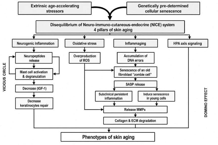
Neuro–immuno–cutaneous interactions in stress-related skin aging.
Extrinsic age-accelerating stressors and genetically programmed cellular senescence affect the Neuro– Immuno–Cutaneous–Endocrine (NICE) system through neurogenic inflammation and oxidative stress, the concept of “inflammaging,” and Hypothalamic–Pituitary–Adrenal (HPA) axis signaling. These interacting pathways lead to increased release of neuropeptides, mast cell activation, production of Reactive Oxygen Species (ROS), cellular senescence, secretion of the Senescence-Associated Secretory Phenotype (SASP), release of matrix metalloproteinases (MMPs), and degradation of the Extracellular Matrix (ECM), all of which collectively contribute to the skin-aging phenotype.
4.5. Hypothalamic–pituitary–adrenal Signaling
Psychological stress activates the Hypothalamic–Pituitary–Adrenal (HPA) axis, a key player in the skin–brain dialogue, leading to the systemic release of glucocorticoids, mainly cortisol. This is adaptive in the short term, but long-term activation burdens cutaneous structure and function. High glucocorticoid levels have been shown to delay barrier recovery after disruption, primarily by interfering with lipid synthesis and lamellar body secretion from the stratum corneum [61]. At the cellular level, cortisol inhibits keratinocyte proliferation and differentiation by promoting epidermal thinning and decreased turnover, and it inhibits fibroblast activity and collagen production in the dermis [62]. This is further enhanced by reduced transforming growth factor-β signaling and increased matrix metalloproteinase expression, which favor extracellular matrix degradation [62]. Importantly, the skin itself contains a peripheral HPA axis (with the ability to locally produce corticotropin-releasing hormone, adrenocorticotropic hormone, and even cortisol, thereby amplifying the stress signal within the cutaneous microenvironment) [63]. These local NE networks indicate that stress-associated aging is not just a systemic process but also a tissue-specific one. Both central and peripheral HPA signaling are chronically dysregulated and are associated with characteristics of premature skin aging, such as decreased elasticity, impaired wound healing, and a higher incidence of inflammatory dermatoses. These findings suggest that HPA axis activity is an important mechanistic connection between psychological stress and structural and functional decline in aging skin, and that therapies that restore neuroendocrine balance may have therapeutic potential.
5. NEUROCUTANEOUS RECEPTORS
The skin harbors a wide variety of distinct receptor types involved in barrier function, immune surveillance, and tissue homeostasis, which guide the direction of aging. While classical receptor systems generally amplify inflammatory signaling and oxidative stress, transient receptor potential channels have a greater influence in this regard, and cannabinoid receptors have a counter-regulatory effect on homeostasis. Serotonin and dopamine receptors also seem to promote cellular resilience, thereby enhancing keratinocyte viability and wound healing [64]. In addition, so-called ectopic receptors in the skin may participate in other physiological functions in regeneration, such as promoting keratinocyte migration and fibroblast activity, as well as modulating inflammation and local tissue tone through acetylcholine receptors. At the translational level, any approach that modulates TRP signaling pathways to reduce activity and/or augment or mimic cannabinoid and/or monoaminergic signaling pathways is a candidate for restoring a more youthful cellular environment. Similarly, selective modulation of ectopic receptors can restore the skin to a reparative and/or quiescent state, thus highlighting their potential role in new skin rejuvenation strategies. The details of these receptors are shown below:
5.1. Transient Receptor Potential (TRP) Channels
These are cutaneous, multimodal, Ca2+-permeable sensory ion channels that act as a neuro-inflammatory hub [65]. They consist of 6 families: TRPA, TRPC, TRPM, TRPML, TRPP, and TRPV. They respond to temperature alterations, UV radiation, and pH changes. Overactivation of TRP induces chronic inflammation and accelerated skin aging [66]. TRP is widely expressed in nerve endings, keratinocytes, melanocytes, and immune cells. It acts as a sensory “Switch” at the nerve ending: a UV sensor, a skin-sensitivity inducer, and an age accelerator. Irritant activation of TRP channels at nerve endings leads to SP release, which exacerbates neurogenic inflammation, induces MMPs, and accelerates skin aging [67]. Neurocosmetics are designed to desensitize TRP channels, reduce inflammation, and restore the skin barrier.
5.2. Cannabinoid Receptors (CB)
Cannabinoid receptors are part of the group of G Protein-Coupled Receptors (GPCRs) [68]. CBs are important in maintaining skin homeostasis, regulating the immune system, and reducing inflammation [69]. The cannabinoid receptors produce endogenous “anti-aging” effects by dampening inflammation, countering free radicals, and improving cell regeneration [70]. Insufficiency decreases the skin's antioxidant activity, disrupts mitochondrial function, increases pro-inflammatory markers, promotes skin aging, thins the skin, and impairs wound healing [71]. This suggests topical products containing cannabinoids are potential anti-aging agents, without the psychotropic properties of oral or inhaled cannabis [72].
5.3. Serotonergic and Dopaminergic Receptors in Skin Aging
The skin is a neurohormonal organ that receives and processes neurotransmitters (such as serotonin and dopamine). These neurotransmitters are crucial but often overlooked in skin aging. Serotonin Receptors, such as 5 HT, are expressed in keratinocytes, fibroblasts, and melanocytes. It acts as a natural antioxidant, aids wound healing, reduces inflammation, and pigmentation [73]. Dopamine and its two receptor types (D1 and D2) signaling maintain skin vitality by regulating cell proliferation, sebum production, and immune response [74]. Dopamine behaves differently in wound healing depending on the receptor type involved. D1 receptor signaling enhances angiogenesis and wound healing, while D2 receptor activation hinders them [75]. Serotonin and dopamine, along with their receptors, decline proportionally with age [76]. This will reduce their protective, anti-inflammatory, and metabolic-regulating effects. “Skin is a mirror of the soul.” Chronic stress depletes these neurotransmitters, accelerating skin aging [77]. Imbalances in serotonin-dopamine pathways are linked to inflammation and pigment disorders. Moreover, manipulating these pathways (such as using serotonin and dopamine antagonists at specific doses) may mimic the effects of calorie restriction on individuals' longevity.
5.4. Olfactory Receptors (ORs)
It is the largest G protein-coupled chemosensory receptor family ectopically expressed in extranasal tissues, including skin cells (melanocytes, fibroblasts, hair follicle epithelial cells, and keratinocytes) [78]. These receptors play a crucial role in various skin functions, from enhancing skin health to accelerating wound healing [79]. OR receptors are highly expressed and promote basal cell proliferation, a process essential for wound healing. It prevents skin aging by suppressing oxidative stress in keratinocytes [80]. It also plays an essential role in fibroblast survival and collagen synthesis [81]. In addition to restoring skin barrier function [82], experimental studies show that Sandalore enhances keratinocyte function in wound healing, whereas the antagonist oxyphenylon inhibits this effect [83].
5.5. Acetylcholine Receptors
Acetylcholine (ACh) receptors are members of the superfamily of neurotransmitter‐gated ion channels. They are expressed in two forms: nicotinic (nAChR) and muscarinic (mAChR). Their natural ligand is the endogenous neurotransmitter acetylcholine (ACh). They classically exist in neural tissues. Recently, extraneuronal localization has been recognized in many tissues, including skin cells [84-86]. Moreover, cells that transiently reside in the skin, such as immune system cells, are involved. Exploration of these receptors is a promising target for managing skin inflammation and aging [84-86]. In neural tissues, ACh released from the nerve terminal fuses with the cell membrane via a SNARE complex. Released ACh binds to nAChR to induce muscle contraction. Wrinkles can be reduced by relaxing facial muscle contractions. This can be achieved by inhibiting SNARE complex formation or by direct binding to nAChRs. The non-neuronal cholinergic system regulates Epidermal Homeostasis, glandular function, and immune response. nAChR agonists suppress UV-induced proinflammatory cytokine expression and oxidative stress, both of which are critically involved in dermal photoaging.
6. NEUROTRANSMITTERS AND NEUROACTIVE AGENTS
Skin aging is not just a structural derangement but a complex neurobiological process. Many neurotransmitters and bioactive molecules mediate this process and can activate, inhibit, or modulate it. Imbalances dysregulate the skin microenvironment and contribute to early skin aging. There are four groups of neurotransmitters: Monoamines (Serotonin, Dopamine, Norepinephrine, Epinephrine, Histamine), Amino acids (GABA, Glutamate, Glycine), Neuropeptides (Substance P, Opioids, Neuropeptide Y), and others (Acetylcholine). They may be excitatory, inhibitory, or modulatory inputs to effectors. The table summarizes the cutaneous neurotransmitters important for skin homeostasis and aging processes [84-86].
The skin is now recognized as a functional neuroendocrine system that can synthesize and respond to neurotransmitters independently of central input. Keratinocytes, melanocytes, and dermal fibroblasts express the enzymatic machinery required for local monoamine production, including serotonin and catecholamines, and environmental stressors such as ultraviolet radiation and oxidative stress can dynamically modulate their levels [87, 88]. This local signaling network appears to fine-tune key processes relevant to aging, including keratinocyte proliferation, melanogenesis, and microvascular regulation. In parallel, acetylcholine, produced within the non-neuronal cholinergic system, acts as a widespread regulator of epidermal differentiation, barrier formation, and immune responses, further supporting the concept that skin neurotransmitters function as local cytotransmitters rather than solely as neural messengers [89]. Such observations suggest that alterations in cutaneous neurotransmitter balance may precede visible structural changes, positioning them as early drivers of age-related dysfunction rather than secondary phenomena.
At the tissue level, neuroactive mediators converge on pathways that define the aging phenotype, particularly chronic low-grade inflammation, extracellular matrix degradation, and impaired repair capacity. Ultraviolet exposure induces matrix metalloproteinases, notably MMP-1, which initiate collagen breakdown and disrupt dermal architecture, a process further amplified by senescent fibroblasts through the release of pro-inflammatory and proteolytic factors [90, 91]. Within this context, neuropeptides such as substance P exhibit dual, context-dependent effects, promoting tissue repair and angiogenesis under physiological conditions but showing diminished efficacy with advancing age [92, 93]. Conversely, histamine has been shown to impair keratinocyte differentiation and weaken barrier integrity, linking neurogenic inflammation to functional decline of the epidermis [94, 95]. Emerging data also point to a protective role for inhibitory neurotransmitters; GABAergic signaling in dermal fibroblasts has been associated with enhanced extracellular matrix synthesis and improved resistance to oxidative stress [96]. Taken together, these findings reinforce the notion that neurotransmitter imbalance contributes directly to the molecular and cellular events underlying cutaneous aging. However, further work is needed to translate these insights into targeted therapeutic strategies. The principal mediators of the Neuro-Immuno-Cutaneous-Endocrine (NICE) network, their physiological roles, and their potential contributions to skin aging are summarized in Table 1.
| Mediators | Primary Source | Physiological Roles | Impact on Skin Aging |
|---|---|---|---|
| Substance P / CGRP | Sensory nerve ending | Mast cell degranulation and immune reaction | Neurogenic inflammation, collagen & ECM breaks & remodeling |
| Serotonin (5-HT) | Systemic and local in keratinocytes | Regulates cell survival and melanogenesis | Skin cell apoptosis, impaired barrier, oxidative stress, and uneven pigmentation. |
| Dopamine | Local and Sympathetic nerve | Regulates pigmentation, stress, and vasoconstriction. | Increases cellular senescence, Irregular melanin synthesis, and barrier disruption. |
| Acetyl choline | Neuron and keratinocytes | Muscle contraction, skin adhesion and differentiation | High activity: dynamic expression lines; low activity: delays barrier repair |
| GABA | Fibroblast | Accelerates cutaneous barrier recovery | Alters muscle tone, shifts inflammation, and decreases collagen, elastin, and hydration. |
| ϐ-endorphin | Keratinocytes, Appendages. | Regulate differentiation, migration, and barrier | Impaired regeneration, reduced barrier function, itch and skin aging |
7. NEUROCOSMETICS AND THE EXPANDING BIOLOGICAL HORIZON OF COSMETIC SCIENCE
The emergence of neurocosmetics marks a conceptual departure in skincare, moving from purely aesthetic, surface-level enhancement of skin as a “tissue” to a holistic, systematic approach that treats skin as a “neuroimmunologic organ” [97]. While traditional cosmetics primarily provide symptomatic management that addresses physical appearance (e.g., epidermal hydration, barrier repair, and visible texture), neurocosmetics interact directly with the skin-brain axis by modulating neurotransmitter release and receptor signaling, thereby addressing stress response and skin homeostasis. As the comparative framework summarizes, this paradigm shift moves beyond structural enhancement toward systemic modulation, aiming to reinforce skin resilience, reduce stress-induced flares, and potentially influence psychocutaneous interactions. The transition reflects a more integrated biological understanding of aging and reactivity, positioning neurocosmetics within a holistic model that bridges dermatologic physiology and neurobiology rather than confining intervention to the epidermal surface alone. Table 2 summarizes the differences between traditional cosmetics and neurocosmetics.
| Features | Traditional Cosmetics | Neurocosmetics |
|---|---|---|
| Primary goal | Symptomatic treatment of aging signs | Systematic modulation of the mechanism of aging |
| Core mechanism | Improve structural integrity | Modulate neurotransmitters and receptor signaling |
| Biological focus | Surface-oriented visible changes | Deep behavioral alteration of the sensory neuron |
| Addressed issue | Treat symptom | Treat system |
| Typical ingredients | Humectant emollients | Neurotransmitter mimickers |
| Scope of impact | Primary physical and aesthetics | Holistic therapy |
| Primary goal | Enhance external appearance | Boost skin behavior, resilience and mood |
| Overstimulation | Irritation | Calm and resilient skin |
8. ANTIAGING NEUROCOSMETICS
Neurocosmetics are an advanced, non-toxic, bioactive skincare category designed to modify skin-brain axis neurons and receptors to mitigate skin aging [98]. These products utilize short-chain biomimetic ingredients that act locally to relax muscles, soothe, or regenerate skin. The first group has “Botox-like” effects that target the neuromuscular junction by inhibiting ACh and reducing facial muscle expression. The second group ameliorates chronic inflammation, collagen breakdown, and skin-barrier impairment induced by local HPA-axis activation by reducing local cortisol levels (a danger signal) and increasing endorphin release (feel-good neuropeptides). The last group improves regeneration by restarting cellular repair through enhanced cellular crosstalk within the skin-brain axis, thereby enhancing collagen, elastin, and ECM synthesis.
8.1. Myo-relaxant to Relax Expression Lines
Wrinkles are the consequence of lifelong muscle contraction. The process begins with acetylcholine release at the neuromuscular junction. SNARE complexes dock ACh-containing vesicles to facilitate their fusion with the neuronal membrane. Released ACh binds to postsynaptic nAChRs, initiating muscle contraction. Several neurotransmitter inhibitor peptides have been designed with sequences that resemble and compete with these key steps (SNAP-25, nAChR, and ion channels). These Botox-like myorelaxant peptides mitigate wrinkle depth in a non-invasive, effective, and safe manner. They satisfy growing consumer demand among injection-averse individuals, younger individuals focusing on prejuvenation, and as a post-Botox treatment to extend treatment results. Although the efficacy of topical Botox-like peptides varies widely based on formulation, concentration, and skin barrier penetration, they can be ranked in descending order as follows: Syn-Ake (Tripeptide-3), Vialox (Pentapeptide-3), XEP-018 (or XEP-030/XEP-018), Snap-8 (Acetyl Octapeptide-3), Argireline (Acetyl Hexapeptide-8), Myoxinol (Hibiscus Esculentus Extract), and Leuphasyl (Pentapeptide-18). Table 3 summarizes the proven Botox-like myorelaxant peptides, in descending order of efficacy, for reducing wrinkle depth after 4 weeks of continuous daily use.
| Name | Ingredient | Source | Year | Developer | Mechanism of Action | Wrinkle Depth Reduction | Ref |
|---|---|---|---|---|---|---|---|
| Syn-Ake | Tripeptide | Snake venom mimic | 2006 | DSM | nAChR antagonist and modulates GABAA receptors | 52% | [99] |
| Vialox | Pentapeptide-3 | Synthetic | 2003 | DSM | Cura-like antagonist of nAChRs | 49% | [100] |
| XEP-30 and XEP-018 | μ-conotoxin CnIIIC McMahon | marine cone snail venom mimic | 2014 | Activen | Blocks ACh release by targeting Voltage-gated sodium channels | 48% | [101] |
| Snap-8 | Acetyl Octapeptide-3 Ji | Synthetic extract | 2003 | Lipotec | Elongated Argireline SNARE destabilizer | 38% | [102] |
| Argireline | Acetyl Hexapeptide-8 | Synthetic extract | 2001 | Lipotec | SNARE destabilizer and blocks ACh release | 30% | [103] |
| Myoxinol | okra extract | Natural extract | 2002 | BASF | Enhancing GABAergic transmission | 26% | [104] |
| Leuphasyl | Pentapeptide-18 | Mimic enkephalin | 2005 | Lipotec | blocking calcium channels | 24% | [105] |
8.2. Neuro-relaxant to Combat Stress-inducing Skin Aging
Chronic stress accelerates skin aging by activating the local HPA axis and the 11β-HSD1 enzyme, which converts inactive cortisone to biologically active cortisol, a “stress hormone,” leading to increased glycation and structural damage of collagen and elastin, catabolic degradation of dermal proteins, and a reduction in the natural moisturizer hyaluronic acid, thereby impairing the skin barrier. The body counteracts this by releasing β-endorphins, a “skin wellness neurotransmitter,” from the degradation of POMC. The Complex β-endorphin/µ-opiate inhibits signal transmission from nociceptors, fine-tunes the immune response, accelerates skin regeneration, and promotes wound healing. Neuro-relaxing anti-aging neurocosmetics ingredients address the “stress-aging” cycle via the skin-brain axis by targeting the local HPA axis, inhibiting the 11β-HSD1 enzyme, or boosting “feel-good ” neurotransmitters, such as endorphins, to improve skin appearance. Table 4 summarizes the proven neuro-relaxant neurocosmetics available commercially.
| Name | Source | Developer | Year | Action | Ref |
|---|---|---|---|---|---|
| Agascalm | Agastache mexicana | Provital | 2015 | Prevents NF-κB release; decreases redness and TEWL; reduces cytokine release | [106] |
| Betaphroline | Tephrosia purpurea (wild indigo) | Givaudan | 2012 | Stimulates β-endorphin, Pain relief, and improvement in well-being | [107] |
| Happybelle-PE | Vitex agnus-castus extract | Mibelle | 2005 | Increases collagen I and III; improves skin hydration and firmness; decreases wrinkle depth. | [108] |
| Happy Skin | Rhodiola rosea root extract | Provital | 2013 | Protection from free-radical-induced damage; Increase in endorphin levels | [109] |
| Neurophroline | seeds of Tephrosia purpurea extract | Givaudan | 2016 | Inhibition of cortisol release; stimulation of β-endorphin production | [110] |
| Sepicalm S WP | Nymphaea alba flower extract | Seppic | 2006 | β-endorphin production increases skin water content and decreases redness. | [111] |
| Nirvanesque | Paeonia suffruticosa extract | NUXE | 2022 | Produces enkephalins as a “neuro-relaxing” molecule | [112] |
| NeuroCalm™ | Peonia suffruticosa | LANCÔME | 2026 | Soothing day cream moisturizes and nourishes | [113] |
| Defensil SOFT | Edible mushroom Albatrellus ovinus | Rahn | 2017 | Blocks TRPV1 receptor; reduces skin hyperalgesia and neurogenic inflammation | [114] |
| Mariliance | Rhodosorus marinus, a red microalga | Givaudan | 2015 | Reduce TRPV1 expression, release of IL-1α, & sensitivity | [114] |
| Neutrazen | synthetic tripeptide | Lucas Meyer Cosmetics | 2000 | Biomimetic of (POMC) reduces SP, IL-1, IL-8, and neurogenic inflammation. | [115] |
| Ocaline PF | Cucurbita pepo Seed Extract | Givaudan | 2025 | Preventive effect on sensitive skin/stinging, soothing of irritated skin | [116] |
| Pinolumin | Swiss stone pine wood | Mibelle | 2016 | Inhibit release of (CGRP, IL-8, and PGE2), act on TRPV1; improvement skin tone | [117] |
| SensAmone P5 | Synthetic pentapeptide from sea anemone | Mibelle | 2017 | Inhibition of TRPV1 receptor activation; reduction of skin sensitivity. | [115] |
| Sensityl | Phaeodactylum tricornutum | Givaudan | 2019 | skin soothing; control of the acute phase of inflammation, immune cell recruitment and inflammatory cytokines | [118] |
| Skinasensyl LS 9749 | Synthetic Acetyl Tetrapeptide-15 | BASF | 2008 | Binding to the µ-opioid receptor; inhibition of CGRP release; increased skin tolerance threshold. | [115] |
| Sepicalm S WP | water lily extract | Seppic | 2006 | production of ß-endorphins; inhibition hyaluronidase; Increase skin water, reduce redness and stinging sensation | [119] |
8.3. Skin Regeneration Boosters
Regenerative skin boosters and neurocosmetics target a biological decline that accelerates after age 50: increased epidermal turnover, fibroblast senescence, declining collagen and elastin levels, and ECM damage. Neurocosmetics rejuvenation boosters are formulated with specialized peptides that help restore skin structure, resilience, and barrier function. They contain signal peptides (which stimulate fibroblasts to increase collagen, elastin, fibronectin, and hyaluronic acid through the TGF-β pathway), carrier peptides (which transport the metal ions copper and manganese, which are involved in wound healing, collagen synthesis, and antioxidant defense), and enzyme-inhibitor peptides (which inhibit MMPs to prevent collagen degradation). Representative regenerative peptides and neurocosmetic ingredients, together with their proposed mechanisms of action, are summarized in Table 5.
| Name | Ingredient | Year | Developer | Mechanism of Action | Ref |
|---|---|---|---|---|---|
| Matrixyl® | Palmitoyl Pentapetide-4 | 2000 | Sederma | Stimulates collagen and ECM components | [120] |
| Decorinyl™ | Tripeptide-10 Citrulline | 2010 | Lipotec SAU | Regulates fibrillogenesis | [121] |
| Biopeptide El™ | Palmitoyl Hexapeptide-12 | 1992 | Sederma | Promotes collagen, elastin and glycosaminoglycans production. | [122] |
| Syn®-Coll | Palmitoyl Tripeptide-5 | 2012 | dsm-firmenich | Boosts collagen production by stimulating TGF-β production | [123] |
| Dermican™ | Acetyl Tetrapeptide 9 | 2011 | BASF | Stimulates collagen type I and lumican synthesis. | [123] |
| Syniorage™ | Acetyl Tetrapeptide-11 | 2013 | BASF | Accelerates keratinocyte growth and syndecan-1 synthesis. | [123] |
| Lipospondin | Elaidoyl Tripeptide-37 | 2010 | Sederma | Stimulates TGF-β, promoting collagen and elastin production. | [123] |
| ND | Peptamide-6 /Hexapeptide-11 | 2012 | Lonza Personal Care | Modulates key genes responsible for collagen synthesis, improving skin elasticity. | [123] |
| ProgelineTM | Trifluoroacetyl Tripeptide-2 | 2012 | Lucas Meyer Cosmetics | Inhibits progerin synthesis, a protein that accelerates cell aging. | [123] |
| Rigin™ | Palmitoyl Tetrapeptide-7 | 2011 | Sederma | Decreases IL-6, stimulates laminins IV and V and collagen VII | [123] |
| Perfection Peptide P7™ | Heptapeptide | 2014 | Mibelle Biochemistry | Stimulating Nrf2-dependent antioxidant enzymes. | [123] |
| GEKG | Tetrapeptide-21 | 2011 | Evonik Industries | Stimulates collagen synthesis at the mRNA and protein levels | [123] |
| KP1 | SA 1-III | 2020 | Università di Firenze | Modulates collagen turnover. | [123] |
| No | Soybean peptides | 2015 | Shenzhen | An enzyme-inhibitor peptide neutralizes free radicals, increases collagen production, and offers UV protection. | [123] |
| PeptAlde™ 4.0 | Rice peptides | 2020 | BASF | Enzyme-inhibiting peptide, Antioxidant, and anti-inflammatory effects. | [123] |
| Activated Silk™ peptides | Silk peptides | 2020s | Nature | An enzyme inhibitor peptide inhibits MMP-1 and stimulates hyaluronan synthase 2 gene expression. | [123] |
| Cu-GHK | Copper Tripeptide-1 | 1990s | Dr. Loren Pickart. | Carrier-regenerative peptide improves healing, modulates MMP-1, promotes collagen and glycosaminoglycans, and has antioxidant properties. | [123] |
| GHK-Mn2+ | Manganese Tripeptide-1 | 1980s | Dr. Loren Pickart. | Carrier-regenerative peptide improves healing, acts as an antioxidant, and reduces hyperpigmentation. | [123] |
9. LIMITATIONS AND KNOWLEDGE GAPS
Other regeneration-based strategies, such as stem cell-derived extracellular vesicles and growth factor-based formulations, could also indirectly stabilize the neuroimmune axis by reducing inflammation and boosting dermal repair. The next step in research should be to assess whether these interventions can alter stress-related signaling networks in senescent skin. The field is conceptually sound, but it has limitations. Multiple studies are in development, but they include small sample sizes or surrogate markers. It is difficult to differentiate expectancy-induced placebo effects from biochemical modulation. Additionally, stressors vary by person, leading to different microbiome compositions; each person's neuroendocrine system also responds differently to stressors, making generalization difficult. Standardized protocols, reproducible biomarkers, and longitudinal designs are required to determine whether modulating the skin–brain axis has long-term benefits.
CONCLUSION
The skin–brain axis is an important framework for conceptualizing the interplay between psychological stress, neuroimmune signaling, oxidative stress, and inflammation in skin ageing. The new generation of neuroactive/regenerative therapies has promise for maintaining skin homeostasis and counteracting stress-induced ageing effects. However, evidence remains limited and inconsistent. More well-designed clinical trials are needed to determine the therapeutic significance of targeting the skin–brain interaction in ageing skin using objective biomarkers.
AUTHORS’ CONTRIBUTIONS
The authors confirm their contributions to the paper as follows: A.T.K, and H.B.: Study conception and design; W.H.Q.A., Y.D.M.Z., and O.Y.A.: Literature collection and data acquisition; A.T.K., H.B., and M.N.S.A.M.: Analysis and interpretation of the literature and findings; A.T.K., W.H.Q.A.: Drafting of the manuscript; A.T.K., H.B., Y.D.M.Z., O.Y.A., M.N.S.A.M.: Critical revision of the manuscript. All authors reviewed the manuscript, approved the final version, and agreed to be accountable for the work.
LIST OF ABBREVIATIONS
| MMP | = Matrix Metalloproteinase |
| ROS | = Reactive Oxidative Stress |
| TRP | = Transient Receptor Potential |
| HPA | = Hypothalamic–Pituitary–Adrenal |
| NICE | = Neuro-Immuno-Cutaneous-Endocrine |
| SASP | = Senescence-Associated Secretory Phenotype |
CONFLICT OF INTEREST
Dr. Ahmad Taha Khalaf is the Editorial Advisory Board member of The Open Dermatology Journal.
ACKNOWLEDGEMENTS
The authors thank Shamil Mahmood Ali Al-Jobore, Al-Salam Teaching Hospital, Mosul, Nineveh Governorate, Iraq, for his valuable support during the revision of the manuscript.